
Invited
Can dynamic force microscopy really "see" intermolecular bonds?
1School of Physics and Astronomy, University of Nottingham, Nottingham NG7 2RD, UK
2Physics Department and Materials Science Institute, Lancaster University, Lancaster, LA1 4YB, UK
3Institute for Interdisciplinary Research Jianghan University Wuhan 430056 China
4Department of Physics, King's College London, Strand, London WC2R 2L, UK
Since Gross et al.'s pioneering observation of the internal "architecture" of a pentacene molecule in 2009 [1], there has been a rapid growth in ultrahigh resolution imaging of single molecules using dynamic force microscopy [2]. Image interpretation is, however, always a challenge with any type of scanning probe technique as, very often, the precise role of the probe can be difficult to 'deconvolve' from the experimental data. I shall discuss a series of ultrahigh vacuum, low temperature force microscopy results (based around the qPlus sensor [3]) for a variety of molecules adsorbed on both metal and semiconductor surfaces: NTCDI [4,5] and biisonicotinic acid (which are both largely planar), and C60 (which is, of course, very far from planar) [6].
Although the biisonicotinic acid data (see Fig. 1) were acquired with a CO-terminated tip, as per the standard approach in the dynamic force microscopy community, high resolution imaging of both NTCDI and C60 involved imaging using a tip which, to the very best of our knowledge, was terminated by an NTCDI or C60 molecule respectively. Intramolecular resolution was possible for tips that interacted both very strongly (~ 2 nN) and very weakly (~ 50 pN) with the Si(111)-(7×7) surface, demonstrating that an inert probe is not a prerequisite for intramolecular imaging. This has important implications for the continuing development of 'internal' imaging of adsorbed molecules on semiconductors.
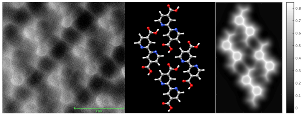
Figure 1: (L-R) Experimental constant height frequency shift image of a monolayer of biisonicotinic acid on Au(111); geometry-optimized structure of the biisonicotinic assembly (determined using the CP2K DFT code [7]); constant height image simulated using the analytical model developed by Hapala et al. [8]
[1] L. Gross et al., Science 325, 1110-1114 (2009)
[2] N. Pavliček and L. Gross, Nature Rev. Chem. 1, 0005 (2017)
[3] F.J. Giessibl, Rev. Mod. Phys. 75 949 (2003)
[4] A. Sweetman et al., Nature Comm. 5, 3931 (2014)
[5] A. Sweetman et al., Phys Rev B 90, 165425 (2014)
[6] S.P. Jarvis et al., Phys Rev B 92, 241405(R) (2015)
[7] https://www.cp2k.org/
[8] P. Hapala et al., Phys Rev B 90, 085421 (2014)