
Invited
Imaging strain waves in a PhotoEmission Electron Microscope: Magnetism goes surfing
1ALBA Synchrotron Light Source, Spain
2Institut de Ciència de Materials de Barcelona (ICMAB-CSIC), Spain
3Dept. of Condensed Matter Physics, University of Barcelona, Spain
4Institut für Physik, Johannes Gutenberg Universität Mainz, Germany
5Swiss Light Source, Paul Scherrer Institut, Switzerland
6Paul-Drude-Institut fur Festkörperelektronik, Germany
Surface Acoustic Waves (SAWs) are propagating Raleigh waves in the upper micrometric layer of a crystal, which propagate large distances and can be generated in piezoelectric materials by applying RF electric fields to transducer electrodes on the surface. We have shown that it is possible to image such waves in LiNbO3 by X-ray PhotoEmission Electron Microscopy (XPEEM), since it is sensitive to the piezoelectric part of the SAW. Thus, the effect of fast strain changes on structures grown on LiNbO3 can be studied with temporal resolution below 100 ps and up to 50 nm spatial resolution, offering a novel approach for studies of dynamic strain in a wide range of research fields (crystallography, nanoparticle manipulation, chemical reactions, magnetism…) The application to the study of dynamic magneto-elastic effects in Ni microstructures will be presented. The magneto-elastic effect or inverse magnetostriction (i.e., the change of magnetic properties caused by an elastic deformation) has attracted much interest due to its potential to write small magnetic elements by an electric voltage rather than current, avoiding energy losses. However, so far practical demonstrations were realized on timescales far too slow for most applications. Combining XPEEM with XMCD magnetic contrast we have imaged the Ni magnetic states at the different phases of the SAW, demonstrating that the strain-driven magnetic anisotropy changes on the sub-nanosecond timescale are as efficient as for the static strain case. Different delays (100-300 ps) of the magnetic response to the strain wave were measured, depending on the magnetic configuration. The delays are related to the intrinsic magnetization dynamics of the Ni patterns. Thus it is shown that while the magneto-elastic effect itself is very fast, for the speed of a potential device the magnetization state has to be properly designed.
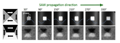
Figure 1: Left, top: Scheme of magnetic domains in a Ni square without net anisotropy, forming a Landau flux closure state (arrows indicate magnetic directions and gray color contrast in XMCD-PEEM). Left, bottom: domain configuration if an additional uniaxial magnetic anisotropy is induced, favoring horizontal magnetization (black and white domains). Right, top: Series of direct images taken with different respective positions of the strain wave and a Ni square (2 µm side). The square is in the maximum of the strain wave (white zone) between the images of 150° and 210° and in the minimum between 330° and 30°. Right, bottom: Corresponding images with XMCD magnetic contrast showing the magnetic domains in the Ni square.
[1] M. Foerster et al., arXiv:1611.02847.